Research

Learn more about the Lubman Lab's work and impact.

Beckman PF2D System
Thermofinnigan Linear Ion Trap
Waters LCT

Lubman Lab Overview

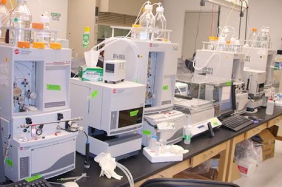
Glycoproteins provide fertile ground for biomarker discovery given their ubiquity in serum, blood and other biofluids. The complex carbohydrate groups that bind to the primary protein may be branched or charged, and these carbohydrates change in their composition and location in the setting of cancer and in other diseases. Analyzing these disease-specific changes can speed diagnosis, ascertain disease stage and progression and help monitor treatment. Our laboratory uses a wide range of methods and technologies – including mass spectrometry, many types of separations methods and antibody and protein microarrays – to analyze and profile large numbers of proteins, including glycoproteins.

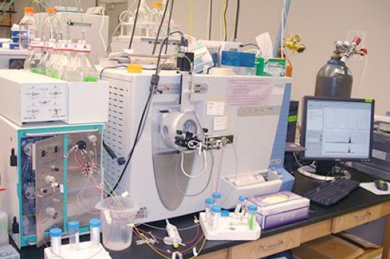
Dr. Lubman and his team have developed a total liquid 2-dimensional separations method based upon isoelectric focusing in the first phase followed by nonporous RP HPLC to generate an image of the protein content of a cell lysate. The proteins in the liquid phase can then be analyzed by electrospray-TOF mass spectrometry to produce a molecular weight map of the proteins in the cell up to 100 kDa.

In addition, tryptic mapping and MALDI-TOF MS can be used to identify proteins against various computer DNA databases. The result is that comparisons can be made between different cancer cell lines to identify proteins that might be important biomarkers of cancer.

Further details on the structure of proteins identified as important to cancer are being determined using sequencing by capillary liquid chromatography/TOF-mass spectrometry, where changes in sequence and posttranslational modifications can be determined. Tissue microarrays are being used to learn about the response of antibodies to these proteins in different tissue samples.

Ultimately, proteins identified as being important to a specific type of cancer are being developed into a protein chip format as a means for early diagnosis of cancer. Biostatistics are being used to determine which proteins are important in cancer transformation and how different cancers can be classified according to the protein expression of the cell.

We investigate how these sugar-coated proteins are differentially expressed by cancer and normal cells. Understanding the ways in which proteins are modified or overexpressed in the context of cancer will provide a greater understanding of the pathways involved in tumorigenesis and metastasis. Extending our work has led us to develop new methods and instruments for two additional frontier areas in the field of proteomics: the detection and analysis of exosomes and of single amino acid variants.

Strategies

The Lubman Lab is tackling the problem of cancer detection from a number of angles. In particular we are working toward identifying biomarkers to enable the use of liquid biopsies. Strategies toward this goal include:

  1. Glycoprotein Analysis in Serum:  This includes quantification and structural changes to identify potential biomarkers.
  2. Isolation & Analysis of Exosomes:  These lipid-coated vesicles are given off and differentially expressed by all types of cells, including cancer cells. Exosomes contain nucleic acids, proteins and glycoproteins, and represent another promising area of biomarker discovery with respect to cancer and other diseases.
  3. Isolation & Analysis of Circulating Tumor Cells (CTCs):  CTCs play a role in cancer metastasis, but their capture and analysis has proven challenging since they occur in very low numbers. Our laboratory works with a commercial partner to filter CTCs from blood so we can analyze them for protein content and surface markers of disease progression, particularly in later stages.
  4. Single Amino Acid Variants (SAAVs):  Common genetic variations in DNA nucleotides, known as SNPS, for single nucleotide polymorphisms, lead to proteins with SAAVs. SAAVs have been implicated in several diseases, including sickle-cell anemia and, we have detected hundreds of SAAVs in cancer cells and in serum.

Our laboratory conducts proteomic analyses of cancer cell lines and cancer cells to identify those SAAVs related to the disease and that potentially may serve as biomarkers.

Results

Instrumentation Development

The Lubman Research Laboratory has made many advances in instrumentation development over the years, including new methods and technologies for detecting, separating and analyzing proteins and glycoproteins. The quadrupole ion trap/time of flight (QIT/TOF) mass spectrometry system and the protein fractionation technology our lab developed for the liquid separation of proteins from cells and blood have been commercialized and being used worldwide. The glycoarray platform we developed has enabled us to generate intact glycoprotein arrays from serum using lectin extraction, and we can use several lectins to study the different responses between glycoproteins from cancer serum versus normal serum or serum in the presence of inflammation.

In addition, we have developed a high-throughput screening assay using an antibody array for these specific proteins followed by a lectin sandwich assay where we can follow the response in large numbers of samples. This can be performed for early stage cancer and for other clinical conditions. The platform has been commercialized and is being used in hospital-related biomarker studies for early detection, prognosis and therapeutic monitoring. Our laboratory also developed the matrix-assisted laser desorption/ionization (MALDI) time-of-flight mass spectrometry technique for the rapid screening of bacteria for changes in protein expression. First published in 1992, the technique continues to be used in both research laboratories and clinical settings today. We continue to generate novel methods for the purification and analysis of exosomes using multiple cycles of centrifugation, ultracentrifugation in combination with other technologies and most recently novel chromatographic techniques. The technologies and methods developed in our laboratory are having an ongoing impact on proteomics and cancer research.

Biomarker Discovery

Our work on the analysis of protein surface markers of cancer stem cells (CSCs) has led us to CD90 as a potential biomarker of glioblastoma CSCs. Although CD90 has been linked to other types of cancer, we are the first to identify its presence in the context of glioblastoma.  We also were the first to identify the glycoprotein haptoglobin as a marker of pancreatic cancer, and as we later discovered, of hepatocellular carcinoma as well. In both cases, changes in the patterns of carbohydrate attachment to the protein peptides have the potential to identify early-stage cancers and differentiate it from other conditions, such as diabetes in the case of the pancreas or cirrhosis in the case of the liver.

Our work has identified other potential markers of pancreatic cancer, including the glycoproteins CD24, RPN2 and CD13, plus a potential marker panel of AACT, THBS1, HPT, and CA 19-9. In addition, we have identified several single amino acid variants, including in the KRAS gene, known to drive pancreatic cancer development, and we have identified several potential serum-based markers and autoantibody arrays for monitoring the response to chemotherapy. In ovarian cancer, we have identified leucine-rich alpha-2-glycoprotein (LRG1) as a potential marker, including in the early stages of disease, alone or to complement CA-125. Our work on exosomes suggests they can be used to detect markers in order to monitor patient response to chemotherapy and radiation treatment. Related to Crohn's disease, we have identified glycoproteomic markers that differentiate intestinal fibrosis from inflammation and can potentially help determine prognosis and monitor treatment response.

Clinical Relevance & Impact

Our laboratory work has and will continue to have significant impact in the clinic. For example, the MALDI technique for high-speed profiling and identification of bacteria developed in our laboratory speeds diagnosis and treatment of life-threatening infections and infectious diseases. The markers we are identifying to detect cancer stem cells in many types of cancers can be correlated with disease progression, helping to better evaluate a patient's prognosis and help guide treatment decisions. Markers, such as haptoglobin, may one day soon help clinicians screen patients at high risk for liver and other types of cancers and detect the disease in its early stages, thereby offering patients a wider range of treatment options to improve survival.

Future Directions

We are excited about the many directions our work is leading, including:

  • New investigations into markers related to the development of Alzheimer's disease for detection and early diagnosis
  • The role of the gut microbiome and cancer treatment
  • The effects of exosomes on bariatric surgery outcomes
  • Exosome content as markers for therapeutic treatment
  • Surface markers of circulating tumor cells and how they define populations of cells that cause metastasis

We continually look for new ways to apply our methods and technologies to improve detection and early diagnosis as well as therapeutic monitoring in cancer and many other diseases. This is a pressing and ongoing need in medicine.

Collaborations

Our work is highly collaborative. We work with many investigators at U-M and other institutions, for example:

  • Neehar Parikh, MD, a liver specialist at U-M, and Amit Singal, MD, of the University of Texas Southwestern Medical Center, on markers of liver cancer and nonalcoholic steatohepatitis.
  • Kyle Cuneo, MD, of the U-M Department of Radiology Oncology, on exosomes as markers in pancreatic cancer.
  • Yehia Mechref, of Texas Tech University, on isoforms of glycans.
  • Lingjun Li, of the University of Wisconsin School of Pharmacy, on structural analysis of glycans using advanced mass spectrometry technologies.
  • Paul Stemmer, of Wayne State Institute of Environmental Health Sciences on single-cell detection of single amino acid variants (SAAVs).
  • Michael Shortreed, in the University of Wisconsin Department of Chemistry, on bioinformatic methods for analyzing SAAVs.
  • Liang Li at the University of Alberta on the metabolomics of exosomes in pancreatic cancer.
  • Haidi Yin, of Hong Kong Polytechnic University, on structural analysis of blood-based glycoproteins.
  • Ryan Stidham, MD, at U-M, on biomarkers of Crohn's disease.
  • Grace Chen, MD, PhD, at U-M, on effects of the gut microbiome on colorectal cancer.
  • Mark Cohen, MD, in the U-M Department of Surgery, on proteomic analysis of head and neck cancer cells lines.
  • Suyu Liu, at MD Anderson Cancer Center, on bioinformatics and biomarker analysis.
  • Liangliang Sun Michigan State University-CZE MS for SAAVs analysis.
  • Haixu Tang, at Indiana University, on new bioinformatic methods for structural analysis of glycans.
  • Tony Hu, at Arizona State University, on microchip-based methods of detecting changes in exosome proteins for chemotherapeutic monitoring.
  • Protein Metrics, on work to better understand the structure of glycans.
  • CelSee, on isolating circulating tumor cells.
  • National Institutes of Health (NIH) National Cancer Institute (NCI) R01:  Serum Glyco-Markers of Early Hepatocellular Cancer
  • National Institutes of Health(NIH) National Cancer Institute(NCI) R01: Universal Internal Standard for Reproducible Accurate Quantification of Exosome Protein Markers
  • NIH NCI U01:  Screening of Glycan Markers in Serum for Early Detection of HCC in Different Etiologies of Disease
  • NIH National Institute of General Medical Sciences R01:  Differential Mapping of Tumor Cells
  • NIH NCI R21:  Therapeutic Monitoring in Pancreatic Cancer Using an Exosome Based Mass Spec Assay
  • The Eli Broad and Edythe Foundation:  Serum Glycoproteome Profiling to Detect Intestinal Fibrosis in Stricturing Crohn’s Disease
  • NIH NCI R50:  Discovery and Validation of Biomarkers for Early Cancer Detection Using Mass Spectrometry