Research

Learn more about the Neuromuscular Lab's work and impact.

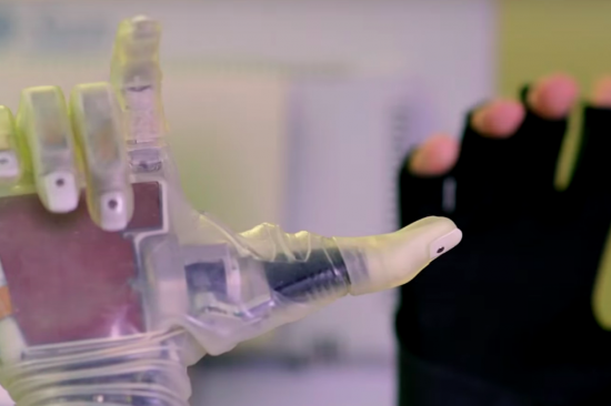
Prosthetic Arm
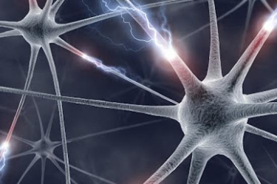
Neuromuscular graphic
Neuromuscular Lab member at microscope

Neuromuscular Lab Overview

Until we understand how to provide prosthetic limbs with intuitive afferent somatosensory feedback — essential for interacting with one’s environment — while simultaneously providing efferent signals for prosthetic control, the ideal prosthetic simply cannot exist. The shortfalls eventually lead to a high degree of abandonment of prosthetic devices, no matter how technologically advanced they may seem. 

Peripheral nerve interfaces for controlling upper-extremity prosthetic devices are not novel, but approaches to date have required electrodes to be placed around or within the nerve itself. However, these interfaces have proven unstable. They often damage the peripheral nerve, and the signal degrades over time. Moreover, they are not effective at preventing neuroma formation or recurrence.

Instead, our approach involves creating a regenerative peripheral nerve interface (RPNI) construct by implanting a residual peripheral nerve into an autologous muscle graft that has been denervated and devascularized. An electrode records signals from the nerve but can be implanted in the muscle, thereby preventing nerve damage. The grafts serve as biologic targets within which the nerve ends can regenerate and signal. In fact, we have observed rapid and robust formation of new neuromuscular connections within the interface in our experiments. As we performed this research, first in small and large animal models and then in humans, we saw that no new neuromas formed in these cases, and we began to investigate the use of RPNIs in both neuroma treatment and prevention.

Strategies

Our laboratory employs many strategies to improve prosthetic control and to both treat and prevent neuromas, including:

  • Development and ongoing investigation of regenerative peripheral nerve interface (RPNI) constructs for prosthetic control
  • Development and ongoing investigation of regenerative dermal sensory nerve interfaces using skin grafts rather than, or in addition to, muscle grafts to provide sensory feedback
  • Development of composite RPNIs (CRPNIs), comprising both sensory and motor nerves, with R21 funding from the National Institutes of Health
  • Investigating the use of RPNIs for treatment of chronic neuroma pain
  • Investigating the use of RPNIs at the time of amputation to prophylactically prevent neuroma formation altogether
  • Use of adipose tissue grafting for enhancing neural regeneration following severe nerve injuries
  • Use of adjacent RPNIs and multiple signals for higher-fidelity motor control
  • Several basic science and clinical studies in these areas are currently underway.

Results

Investigators in our laboratory and our collaborators have demonstrated and continue to characterize the viability and reliability of regenerative peripheral nerve interfaces (RPNIs), as well as composite RPNIs that include sensory nerves and dermal grafts, for prosthetic control.

Our constructs initially used muscle-forming cells, or myoblasts, but our later work has shown greater success with skeletal muscle grafts. These grafts have had their nerves and vasculature removed and, in experiments with animal models, we found that nerve and blood vessel regeneration begins to take place in four weeks. We’ve obtained similar findings in both motor and sensory nerve constructs.

Our patients who have undergone RPNI surgery have achieved greater fine finger control of prosthetic hands, including the simultaneous and independent use of two fingers through multiple, adjacent RPNIs, and the ability to eliminate signal cross-talk among them. We’ve recently obtained funding from the U.S. Department of Defense Defense Advanced Research Projects Agency and an investigational device exemption from the U.S. Food and Drug Administration to continue this work with greater numbers of RPNIs in our patients.

As our work to demonstrate the viability of RPNIs to improve the control of prosthetic devices, we’ve discovered the beneficial effect of RPNIs on neuroma treatment and prevention.

A pilot study conducted among 16 patients found that RPNI constructs reduced their phantom limb pain by more than 50% and neuroma pain by more than 70%. Over half of our patients said they were able to lessen their use of pain medication as a result.

We also have been investigating the use of RPNIs for primary prevention of neuromas. To date, we’ve implanted RPNIs on over 50 patients – including multiple RPNIs on some patients, totaling over 180 implants – at the time of amputation. After a year, we found no evidence of painful neuromas in our patients.

Overall, in close to 200 RPNI procedures completed to date in humans, our patients have not had a single neuroma recurrence.

Clinical Relevance & Impact

The surgical procedure we’ve pioneered to construct and implant RPNIs – as well as the prophylactic use of RPNIs to prevent the formation of neuromas – is now becoming the standard of care at the University of Michigan. 

We expect it to become adopted at other institutions as well since it can be performed at the time of amputation in about 15 minutes by physicians across a broad range of surgical specialties, including orthopedics, trauma, and plastic surgery. 

In addition to treating and preventing neuromas, we can now provide our patients with an interface that will allow them to wear and control a prosthetic device and that can potentially feel what they’re doing, with significantly less pain and discomfort – holding their children, preparing food, raising a glass and many other activities.

Helping patients improve control of prosthetic devices and reducing their pain levels has far-reaching public health and societal impacts, too – less use and potential abuse of opioid medications, lower rates of depression and suicide, lower healthcare costs and improved productivity. We all benefit from this surgical advancement.

Future Directions

Our research continues to lead us to new questions and areas of inquiry. Our work to better understand fat grafting and the mechanisms underlying its role in nerve regeneration continue. We are also looking toward how GABA receptors may influence major depression that often results from chronic pain associated with severe nerve injury, and toward a better understanding of sexual dimorphism and pain perception. Learning how signaling processes differ can guide tailored and more effective treatment approaches. The rat model of neuroma our lab developed for our RPNI work is clinically relevant in this area as well, and we recently received a pilot grant for research that has already begun to generate data.

Collaborations

Our lab works collaboratively across many disciplines, and we are continually expanding the scope of our work to bring new perspectives and insights to our team. Collaborative research efforts range from the neural influence on the development of heterotrophic ossification and bone fracture to new ways to stimulate nerves of the autonomic system to cure or prevent diseases such as type 2 diabetes. Key collaborators on these projects and others include:

  • Department of Defense Peer-Reviewed Orthopaedic Research Program for the Office of the Congressionally Directed Medical Research Programs: Regenerative Peripheral Nerve Interfacesfor the Treatment of Painful Neuromas in Major Limb Amputees
  • National Institute of Health (NIH), R21: Regenerative Peripheral Nerve Interfaces Using Composite Free Muscle and Dermal Constructs
  • American Society for Surgery of the Hand: High-Fidelity, Multichannel Carbon Fiber Electrodes to Enhance Nerve Regeneration
  • National Institute of Health (NIH), SPARC: Highly Compliant Microneedle Arrays for Peripheral Nerve Mapping
  • Plastic Surgery Educational Foundation: Multichannel Carbon Fiber Electrodes to Enhance Nerve Regeneration
  • Space and Naval Warfare Systems Center, Pacific: Providing Intuitive Prosthetic Movement and Sensation Using Residual Nerve Endings to Neurotize Regenerative Muscle Grafts