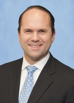
Michael Englesbe, M.D. (Residency 2004), remembers it as a “clear eureka moment.”
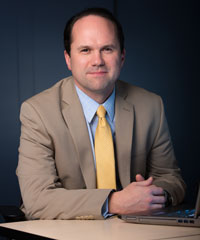
Christopher Sonnenday, M.D., calls it a “convenient kind of coincidence.”
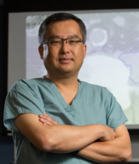
They’re referring to a grand rounds presentation by Stewart Wang, M.D., Ph.D., Endowed Professor of Burn Surgery and professor of surgery, that both attended about two years ago, in which he presented a novel application for the wealth of information contained in patients’ CT scans.
A busy trauma and burn surgeon known internationally for his many years of work with the auto industry to improve vehicle safety and lessen the severity of crash injuries, Wang described a system for analyzing data from accident victims’ scans to determine why some of them fared so much better than others in comparable scenarios.
“Typically,” Wang says, “we describe a victim in a car crash the same way we describe a patient in medicine: age, gender, height, weight, and sometimes that they’re overweight or have an existing medical condition.” But the engineers who consulted him kept pressing for greater precision. Coming from a family of engineers, Wang understood perfectly.
“The only way I could think to answer their questions was to use CT scans,” he says, “so we developed a systematic way of indexing thousands upon thousands of very detailed body measurements. And darn if it didn’t turn out that these were the best explainers for severity and type of injury suffered in a crash. At my grand rounds, I posited that our technique provided a much more granular way of describing physical health and the cumulative effects of disease within each individual patient, and that by better assessing each individual patient’s body, we could provide more personalized and more effective treatment.”
Predictive power for better outcomes
For Sonnenday and Englesbe, both transplant surgeons with a passion for improving outcomes, Wang more than merely posited; he switched on some light bulbs and lit a few fires.
“Mike Englesbe and I, in hearing Stu talk about this, realized some overlap with an area of interest of ours,” says Sonnenday, an assistant professor of surgery in the Medical School and of health management and policy in the School of Public Health. For him, it was how to get smarter about selecting the most appropriate patients for transplant. For Englesbe, an associate professor of surgery, it was how to determine which patients undergoing major surgical procedures were more likely to recover than others. In fact, Sonnenday had just started a large prospective study looking at whether frailty could be measured in candidates for liver transplant, “when I stumbled across this other way to measure physiological health,” he says.
The opportunity was almost too good to be true. Virtually every surgical patient has had a CT scan, and outcomes for liver transplants are unambiguous. “We know how these patients have done, and we already have their imagery, so we can link the two together, so to speak,” says Englesbe.
The key driver of successful outcomes is a surgeon’s ability to pick the patients that are going to do the best, according to Sonnenday. “An experienced surgeon will do an eyeball test to see if a patient looks fit enough to undergo a major procedure,” he says. “My thought was perhaps all this additional information that’s sitting there on these images actually is a way to objectify the eyeball test.”
Instead of just relying on one’s surgeon’s judgment and experience, actual data could be communicated between providers and between centers, and explained to patients to educate them on what exactly their risk may be. “Could we quantify things from these images that would tell us something about the overall health of the patient and their fitness for surgery?” he wondered.
It’s not stretching the metaphor to say that what Sonnenday calls “our first pass at that” was completed for a touchdown. They found 200 liver transplant patients who had CT scans right before or right after their operations, and correlated their outcomes with the size of their psoas muscles.
“We kind of picked it out of a hat, to be honest, because it would be easy to measure,” says Sonnenday. The psoas runs along the spine and into the thighs. Not only is it a core muscle, it’s also relatively easy to distinguish from its surroundings.
“You can pretty easily teach a computer an algorithm to find the psoas muscles and measure their size and density, so that’s what we did,” he says. “Dr. Wang’s lab had already exerted a lot of effort in how to measure these things; they’d just never been used for a clinical purpose before.”
What they found in that first study was that psoas muscle size correlated extremely well, to say the least, with liver transplantation survival. Simply put, the bigger the psoas muscle, the better the patient did. The magnitude of the effect overwhelmed the statistical model, linking more strongly with outcomes than any of the other traditional indicators like characteristics of the recipient, age, diagnosis, other illnesses, or characteristics of the donor organ.
“That kind of got us thinking we were on to something,” says Sonnenday.
Subsequent studies correlating other markers — such as bone density, vascular calcification, fat distribution, and chest and abdominal musculature — with outcomes for aneurism and cancer surgery have confirmed their suspicions. Wang points out that a study of patients coming into the ICU for extracorporeal membrane oxygenation (ECMO) showed that muscle size is a better predictor of who’s going to live or die than Apache III, a complex scoring system that’s been used for decades to assess mortality risk.
But, as Sonnenday puts it, “Yes or no on surgery doesn’t really help patients, other than excluding some. If we had a way to improve the candidacy of patients for major medical interventions, that would be a big step forward.”
He, Englesbe and Wang have dubbed what they do with CT scans “analytic morphomics,” one of several terms they’ve had to invent because what they’re up to is too new to have a nomenclature. They call improving patients’ candidacy for surgery “prehabilitation,” and it involves reducing “remedial risk,” a phrase borrowed from the financial world to describe the factors, unlike age or co-morbidities, that patients have the power to alter.
“Analytic morphomics has helped us understand why some people do really well after surgery, and we’ve taken that insight to design ways to better prepare patients for surgery,” says Englesbe. “Two hours of general anesthesia with major abdominal surgery is about as hard on a patient as running a 5K as fast as they can. If we can better prepare them, they can do better. The paradigm shift is that the surgeon’s schedule shouldn’t drive when the operation happens; it should happen when the patient is best prepared for it.”
The first formal realization of that vision is the recently launched Michigan Surgical and Health Optimization Program within University Hospital’s preoperative clinic, which Englesbe directs. The program’s two principal components are risk stratification — helping patients and surgeons make better decisions about whether or not they should have major surgery — and optimization, which prepares patients as well as possible for surgery.
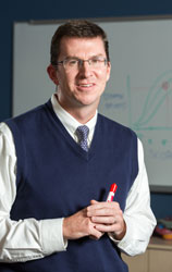
Optimization, says Englesbe, consists mostly of approaches like smoking cessation, better nutrition, walking, strength training, stress reduction, bad habit management and getting family and friends on board to take care of you. An outstanding educator who won the Kaiser-Permanente Award for Excellence in Teaching last summer, Englesbe knows a teachable moment when he sees one.
“Not to sound boastful or flippant, but patients do everything I tell them to do,” he says. “The only people who can say that are surgeons who do major operations on really scared people. It’s a unique opportunity. We see 16,000 patients a year in the pre-op unit, and the whole concept of optimization is relevant to every one of them."
“How it’s applied is between the patient and the clinical experts in the program,” Englesbe adds. “But even with the ideal patient — the young, fit nonsmoker — there’s still a lot that can be done with respect to education and stress reduction, surgical planning with family and friends, missed work, and a lot of the other things that stress people out when they have a health problem. It goes beyond just getting people in shape. It’s really more of a holistic approach that can benefit anyone having surgery.”
And even people who aren’t.
As surgeons, Englesbe and Sonnenday primarily have been concerned with surgical interventions, but as their colleagues have learned about their work and bought into the validity of the science, some of them are now considering its use in other clinical scenarios.
“We’re just starting to look at whether analytic morphomics can help chemotherapy docs titrate chemotherapy,” says Englesbe, “which in some ways is an even more stressful clinical intervention. It seems to predict outcomes there, too.”
How old are you?
Yet another new term illustrates yet another new way to deploy the information lurking in CT scans.
“Instead of looking at imaging to tell us about pathology, we’re looking at imaging to tell us about the patient,” says Sonnenday. “Good clinicians have probably done it for a long time but there’s never been a metric to quantify that. To make it even more tangible, one of the things we’ve been working on recently is the concept of morphometric age.”
The idea behind the concept is easy to grasp. For example, one patient might be an 80-year-old who walks three miles a day and lives independently and is very robust. Another might be a 60-year-old who needs help bathing and has no muscle mass. The 80-year-old is arguably a better surgical candidate than the 60-year-old, even though all the traditional metrics would predict that the 80-year-old would have a higher mortality risk.
“What if we could be smarter than that?” says Sonnenday. “What if, at the bottom of the CT report, some sort of scoring system based on analytic morphomics tells you the morphometric age of that 80-year-old patient is 56? We’re still a ways off, but that’s the type of information we’re trying to pull out of this data.”
“The beauty of morphometric age is that it’s a very intuitive, really patient-centered way to describe surgical risk,” says Englesbe. “If I’m 70 but inside I’m 50, trying to cure my cancer with a big operation seems like a reasonable approach. If I’m 70 but inside I’m 95, it sounds like something less invasive than surgery might be better for me.”
Helping patients understand and evaluate the risks and benefits of surgery is “a really hard conversation to have,” he says. “It’s well accepted that the paternalistic approach is not the best way to come to clinical decisions, but 95 percent of my patients do what I think is best for them. That’s not the way we should be doing it. I try to do research that I can explain to my mother, and this is an approach to an important clinical problem that’s easy to understand and helps patients feel in charge of their care.”
In some respects, the trajectory of analytic morphomics resembles that of genomics. It has the capability of both touching numerous aspects of treatment and rendering it vastly more personalized, but it is still in its infancy. As Sonnenday says, “Our ability to generate the data is ahead of our ability to analyze it and correlate it with clinical outcomes.”
On the other hand, imaging data is available for far more patients than is genetic data, and its use is already well established in clinical practice. Morphomics may yet prove as potent as genomics in individualizing patient care.
“The first thing they teach you in medical school is treat the patient first and foremost, and not just the disease,” says Wang. “But if you look at real practice, most of the time we treat the disease, making some minor modifications for the patient. That’s why there’s this whole investment in the human genome. If you understand the genetic makeup, you better understand the patient and can personalize their treatment.”
In his view, CT scans are just as rich. “What we have is the catalogue of the patient’s body at a point in time,” he says. “We’re using the same high-throughput statistical systems that were developed for genomics to analyze this data and what its relationship is to patient outcomes. We know if we can tailor the treatment, the patient will do better.”
By inspiring a creative way to deploy digitized data living in the clouds, the convergence of the trauma burn surgeon and the transplant surgeons, in real time and physical space, may have spawned an approach to surgery that will dramatically improve outcomes.
Wang finds that kind of synergy, however serendipitous, immensely satisfying.
“The way I was raised was that solving problems is a main reason for your existence,” he says, “and that’s what I love about being an academic here. I love my clinical practice and taking care of patients, but working at the university gives me the privilege of taking some time to work with very smart people to solve problems.”
This story, written by Jeff Mortimer, originally appeared in Medicine at Michigan magazine. Photos by Eric Bronson and Austin Thomason, Michigan Photography. Photo-Illustration by C.J. Burton.

Stewart C. Wang, MD, PhD, FACS
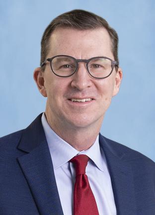
Michael J. Englesbe, MD, FACS
