For Patients
Administrative Contact
Wendy Mrdjenovich: 734-232-8404
Biography
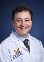
Dr. Miller completed his undergraduate degree in biology at Stanford University, where he was active in development of microsurgical devices with the Department of Ophthalmology under the mentorship of Daniel Palanker, PhD and Mark Blumenkranz, MD. He completed his MD and PhD graduate work at University of California, San Francisco (UCSF), where he developed primary neuronal culture models of Huntington's disease under the mentorship of Steve Finkbeiner at the UCSF-affiliated Gladstone Institute of Neurological Disease. He completed his internal medicine internship in the Bay Area at Kaiser Permanente Oakland. Seeking to combine his post-graduate medical and scientific training, he became the inaugural awardee for the University of Michigan Kellogg Eye Center's Pre-Residency Research Fellowship. This fellowship allowed for establishment of an independent research program prior to joining the ophthalmology residency. He completed the fellowship in 2016, joining the residency program in July 2016 and finishing in June 2019. He then completed a medical retina and research fellowship also at the Kellogg Eye Center before joining the faculty in summer of 2021. His research program seeks to establish primary and iPS RPE culture models of dry AMD as a platform for testing therapeutic interventions. Numerous projects exist in the lab for interested residents, medical students, or other trainees.
Outside of work, Dr. Miller enjoys squash (co-founding the Stanford Squash intercollegiate program while an undergraduate), foreign policy debates (co-founding a national organization dedicated to addressing human rights violations in Sudan), cooking with his wife, and watching his three young children grow up.
Areas of Interest
Research Summary
Human primary and induced pluripotent stem cell-derived (iPSC) RPE culture models of dry macular degeneration; autophagy; RPE lipid biology and metabolism
Age-related macular degeneration (AMD) is the leading cause of blindness in the developed world. The disease comes in two forms - a slowly progressive degeneration called dry AMD and a much more rapidly progressive degeneration involving abnormal blood vessel growth called wet AMD. Starting in approximately 2005, anti-blood vessel medications have led to a radical improvement in our ability to save vision in wet AMD patients. However, for the 80+% of patients with dry AMD, we lack any proven therapeutic intervention. We study the mechanisms driving dry AMD with the goal of identifying therapeutics for this disease.
Macular degeneration affects the retina, the layers of cells in the back of the eye that turn light into electrical signals that the brain can interpret. Humans and primates are unique in the structure and properties of their retina, and it has therefore been difficult to establish non-primate animal models of AMD that truly recapitulate all features of the disease. We therefore seek to use cells that we collect from human retinas to build a model of AMD in a culture dish. The cells most affected in macular degeneration come from a pigmented layer of the retina called the retinal pigment epithelium (RPE). Our experiments involve stressing human RPE grown in the lab with a range of insults (including the insult of just carrying out routine daily activities but over a prolonged period of time), testing whether the RPE responds to the stress in a way that looks like the human disease. In particular, the cells on top of the RPE, called photoreceptors, undergo a daily shedding of their cell tips. These jettisoned fatty debris are cleared each day by the RPE. At the same time, lipid complexes enter the RPE from a set of tiny blood vessels underneath the RPE, called the choroid. These two lipid sources, photoreceptor tips and lipid particles from the blood stream, are an enormous burden to the RPE. Indeed, when the RPE loses its ability to efficiently clear this lipid load, the debris can accumulate outside the RPE as "cellular trash," a first sign of dry AMD. This trash is called "drusen."
In our model system, we feed our RPE cultures photoreceptor cell tips +/- fats that mimic lipid coming from the choroidal vessels, and track how the RPE successfully and unsuccessfully handles this fatty cargo. We are also manipulating RPE pathways involved in metabolism and cellular trash removal to improve the RPE's ability to handle photoreceptor cell tips or toxic debris. These studies have identified drugs which decrease drusen accumulation, which have promising therapeutic potential in dry AMD. Visit the Jason Miller lab website to learn more about our research.
Clinical Interests
- Age-related macular degeneration
- Inherited macular degenerations
- Other macular disease, including central serous choroidopathy, macular telangiectasia, macular toxicity from medications, and macular vascular disorders
- Initial diagnosis of infectious and non-infectious uveitis
- Distinguishing retinal from neuro-ophthalmologic conditions
- Non-surgical management of retinal detachment (pneumatic retinopexy)
Subspecialty: Retina
Make an Appointment
- Patients
- Make an appointment with Dr. Miller
- Information for Patients - locations, maps and directions, patient resources
- Clinical Service: Retina and Uveitis
- Referring Physicians
- M-LINE, 800-962-3555
- Information for Health Providers
In the News

$11.5 Million Gift Fuels Innovation in Dry Age-Related Macular Degeneration Research
Jason Miller, M.D., Ph.D., named first James Grosfeld Professor of Ophthalmology and Visual Sciences and will play key role in new initiative.
Credentials
- MD - University of California, San Francisco
- PhD - University of California, San Francisco
- Residency - Ophthalmology, Kellogg Eye Center, University of Michigan
- Fellowship - Medical Retina, Kellogg Eye Center, University of Michigan
- Postdoctoral Fellow, Kellogg Eye Center, University of Michigan

