Pediatric Cardiac MR

8 year old with double aortic arch on cardiac MRI
Cardiovascular Magnetic resonance imaging (MRI) in children is a valuable noninvasive technique that is used to create detailed images of the heart and vascular structures without radiation risks. We perform cardiovascular MRIs to diagnose and direct intervention in a wide range of heart conditions. Our expert team of faculty and staff is adept in using the latest advances in MR imaging as well as in making the necessary technical adjustments for imaging patients of all ages ranging from small babies to adults.
Our cardiovascular MRIs are performed in the C.S. Mott Children's Hospital at the University of Michigan Health System
Transcatheter Aortic and Mitral Valve Replacement Programs (TAVR/TMVR):

Three-dimensional CT rendering of a TAVR valve (red) in normal position after placement. The white areas are calcium from the patient’s stenotic aortic valve that have been pushed aside by the TAVR valve.

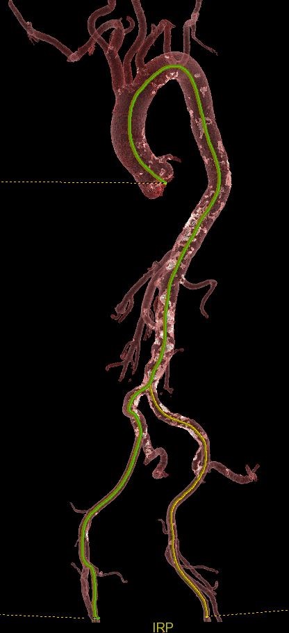
Three-dimensional CT rendering of the aorta and its major branches with “road maps” (green and yellow lines) depicting the planned route for the TAVR valve to get from the legs to the heart.
Patients with severe dysfunction of the aortic or mitral valve, who are candidates for transcatheter replacement of the aortic (TAVR) or mitral (TMVR) valves, undergo a pre-procedural imaging with computed tomography angiography (CTA). Imaging is performed using IV contrast to highlight the heart and blood vessels, and all scans are performed with ECG-gating, a technique that removes pulsation of the heart and creates motion-free images. Pre-procedural CTA is a critical planning step prior to successful transcatheter valve replacement and CTA images are used to determine the appropriate size and fit of the replacement valve and to create a “road map” of the blood vessels to ensure a clear path though which the valve can travel from the groin to the heart. Image analysis experts in the Department of Radiology’s 3D Imaging Laboratory use CTA images to create three-dimensional models and perform detailed measurements of the heart and blood vessels. TAVR/TMVR studies are interpreted by Cardiothoracic Radiologists with expertise in Cardiovascular Imaging, in a close collaboration with a team of cardiac surgeons, cardiologists and interventional cardiologists to ensure the safest and most effective procedure for the patient.
