While brain cancers are not always lethal, the most common form, glioblastoma, is aggressive and incurable, with a median survival of one and a half years. From identifying deadly tumors sooner, understanding the biology of how and why they grow, and uncovering promising new treatments, researchers at University of Michigan Health Rogel Cancer Center(link is external) are carving a path that creates hope for patients.
Two-pronged attack for high grade gliomas
Maria Castro, Ph.D.(link is external), and Pedro Lowenstein, M.D., Ph.D.(link is external), are leaders in tackling aggressive brain tumors. They specialize in gene therapy-mediated immunotherapy for high grade gliomas and collaborate with researchers across U-M to translate their therapies for adult and pediatric patients. Their work, more than two decades in the making, is about to pay off.
“Gliomas are very aggressive, and it’s important to make progress, but that progress can be terribly slow,” says Castro, R.C. Schneider Professor of Neurosurgery, professor of cell and developmental biology and co-director of the Biosciences Initiative in Brain Cancer Technologies(link is external).
“There’s been a lot of progress in identifying gliomas, imaging them and running diagnostics, but this has had little impact on therapeutic outcomes.”
When Castro first began working with Lowenstein on brain tumors in the 1990s, gene therapy was a new, emerging field, and their team was at the frontier’s edge, focused on treating high grade gliomas.
“No one was doing gene therapy,” Castro says. “It was a very new technology.”
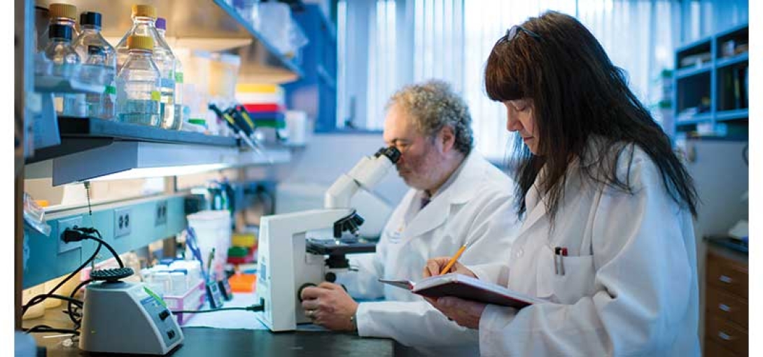
Maria Castro, Ph.D. and Pedro Lowenstein, M.D., Ph.D., are leading researchers on tacking aggressive brain tumors. Credit: Michigan Photography
Single-vector approaches were effective for killing rapidly dividing cancer cells, but sparking an immune response in the brain was challenging. Lowenstein, Richard C. Schneider Collegiate Professor of Neurosurgery, professor of cell and developmental biology, and professor of biomedical engineering discovered that the brain’s immune system lacks dendritic cells, which are crucial for initiating immune responses. That lack could explain the difficulty in eliciting effective anti-glioma immune responses.
To trigger a natural immune response in the tumor cavity after resection, Castro and Lowenstein wanted to use a new two vector approach: “One gene kills the tumor cells, the other activates the immune system,” Castro says. “It’s a two pronged attack.”
Castro, Lowenstein, and their team built two adenoviral vectors to be injected into the brain cavity post-resection. The early results were surprisingly promising, Castro and Lowenstein recall.
“When you use each therapy independently, neither one works so well,” Castro says. “But when you combine them, the efficacy just rises through the roof.”
The therapy also worked particularly well for model animals with gliomas that carried the IDH1 mutation, which Castro describes as “a death sentence delayed in time.” Mutant-IDH1 tumors always come back and are resistant to radiation treatments. But patients with mIDH1 tumors tend to have longer survival times—in part, as Castro recently published in Science Advances, because the mIDH1 tumor microenvironment is epigenetically modified such that the immunosuppressive cells have lost their immune suppressive capacity. Castro and Lowenstein’s dual-vector approach takes advantage of that mutation.
In early studies, they found 98% of animals with mIDH1 glioblastomas were cured, a finding “never seen before in any tumor type,” Castro says. “It worked beautifully.”
Castro and Lowenstein moved to Michigan Medicine in 2011 and began the study of the dual vector treatment, targeting high grade glioma patients with wild type IDH1 in 2013 with support from the Phase One Foundation. The results, published in The Lancet Oncology, from 18 patients treated as part of a Phase I trial were stunning.
Patients in the study saw median survival times of about 21 months, a significant gain from the historic survival of 14.6 months. The two year survival rate was nearly 40%; seven patients survived beyond two years, three survived beyond three years, and one was still alive 60 months after initial study enrollment.
“We believe this gene therapy approach may be even more powerful in patients with the IDH1 mutation,” Castro says. “I predict it will be really effective in that patient population, so that’s one of the future trials we’re working on now.”
Part of the promise of the treatment is the potential for long term therapy. Castro and Lowenstein found that HSV1-TK persisted in patients’ brains for more than two years. Currently, Valacyclovir is only authorized to be administered for a total period of four weeks, so their findings suggest that if the administration of Valacyclovir could be extended to months or years, patient survival times could be extended proportionally.
“This was totally unexpected,” Lowenstein says of HSV1-TK’s longevity. “It gets me particularly excited, it’s very promising. It certainly justifies going into larger trials.”
Lowenstein and Castro are now developing a phase Ib/II trial for the dual-vector approach for wild type and mIDH1 glioma patients. Overall, they hope their work will help the field and that the FDA will have broader acceptance of multi-pronged treatments like theirs.
“The way to push this forward is in combination,” Castro says.
Not only one diagnosis
Pediatric oncologist Andrea Franson, M.D.(link is external), works with Castro and Lowenstein to bring their dual vector treatment to pediatric patients. Franson’s focus has been on high-grade gliomas with the H3G34R mutation, which is present in a subset of patients with pediatric high-grade glioma patients. This effort is supported by Ian’s Friends Foundation(link is external).
Typically, these patients live only 12 to 24 months from diagnosis.
“In so many of these tumor types, the need for better treatments and outcomes is so desperately high,” Franson says. “We’re dealing with these really rare tumors, so thankfully the numbers of patients are low, but there are still a lot of tumors and a lot of need.”
Like mIDH1 tumors, pediatric H3G34R mutant tumors have incapacitated immunosuppressive cells, making them potentially susceptible to the dual-vector treatment. Franson, assistant professor of pediatrics, is working under the mentorship or Castro and Lowenstein to explore anti-tumor immune responses in H3G34-mutated versus non-H3G34-mutated tumors in mice, probing the system for points that could be exploited to trigger an immune attack using the dual-vector treatment.
Translating Castro and Lowenstein’s genetic immunotherapy approach for pediatric patients is critical, in part because so many tried and true treatments have fallen short.
“Chemotherapy, radiation therapy and surgery may work for the short term, but the tumors don’t stay away,” Franson says. “People have tried many, many things over the last several decades, but for the toughest tumors, there’s really been no progress.”
Part of the delay in successfully treating H3G34R high-grade gliomas lies in researchers’ lack of knowledge about their fundamental biology. Scientific understanding of brain tumors’ genetics (including histone mutations like H3G34R) didn’t come about until recent years.
“The more we dig into tumor genetics, genomics and proteomics, the more we realize there are all these subcategories of tumors,” Franson says. “We may be giving patients the ‘same’ diagnosis, but really, it’s not.”
Franson hopes to hone treatment by improving tumor profiling at the diagnostic state. While that reality may be distant, methylation profiling is a promising step down that pathway and may lead to different treatments choices made from the time of diagnosis. Franson is a co-lead investigator for a national a phase I clinical trial that’s using genetic tumor profiling to optimize drug treatment.
“If we understand enough about how these tumors form and progress, we’ll get closer to finding what actually helps patients,” Franson says.
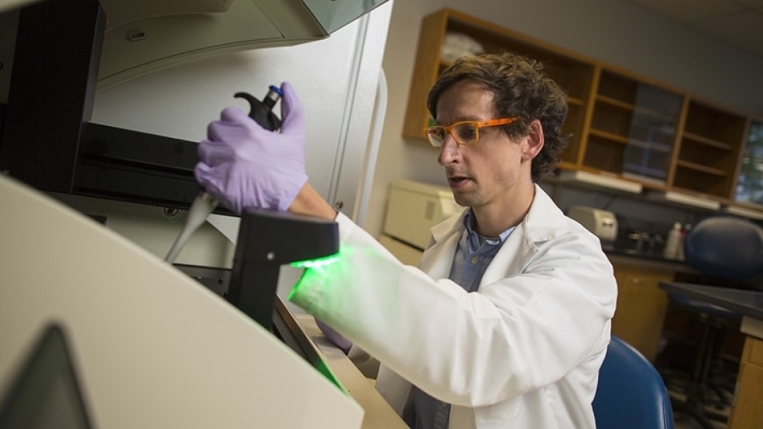
Carl Koschmann, M.D., works in his lab. Credit: Michigan Medicine
Pediatric oncologist Carl Koschmann, M.D.(link is external), who serves as co-director of the Chad Carr Pediatric Brain Tumor Center, is developing a treatment for pediatric patients with diffuse midline gliomas (DMG) with the H3K27M mutation.
The median survival time for patients with these high-grade gliomas is around 12 months, even with radiation therapy, “so there’s a lot of progress to be made,” Koschmann says. “Currently, we can’t remove them. We can barely biopsy them.”
Koschmann’s dual life in the clinic and the lab, including starting his own lab in 2016 after training with Castro and Lowenstein, has enabled him to work on the cutting edge of both understanding and treating tumors with this mutation.
His most recent advancement builds on studies of ONC201, a dopamine receptor antagonist, that has been explored for use in diffuse midline glioma (DMG) patients. In an early study, ONC201 was found to be safe and cross the blood-brain barrier, but it was not effective at treating adult glioblastoma. But several younger patients in that trial who had DMG that harbored a hallmark H3K27M mutation showed a potential response, opening the door to further study.
A phase I trial of ONC201 opened in 2017 to examine the H3K27M mutation and its tie to better survival times. U-M was one of a few early trial sites. During this time, Koschmann worked with Sriram Venneti, M.D., Ph.D.(link is external), the co-director of the Chad Carr Pediatric Brain Tumor Center, to understand why the drug might be more effective in cells with the H3K27M mutation.
Their studies of spinal fluid from patients treated at U-M led to the discovery that ONC201 disrupts mitochondrial behavior and changes the tumor’s epigenetics, “essentially restoring tumor cells to be a lot more like normal brain cells after treatment,” Koschmann says.
Their study, published in Cancer Discovery, shows that in a cohort of 36 DMG patients treated after initial radiation, the median survival time was nearly 22 months. This therapy is the first single therapy to improve outcomes for these patients, and a phase III trial is underway.
“Since 2017, we’ve treated more than 100 patients with this therapy here at U-M,” Koschmann says. “Their overall survival is noticeably better than historical survival, so it’s exciting to see that there is a crack in the armor. I’m optimistic that in our generation of experimental treatments, we’ll have patients surviving five or 10 years, or beyond. That’s where the future is heading.”